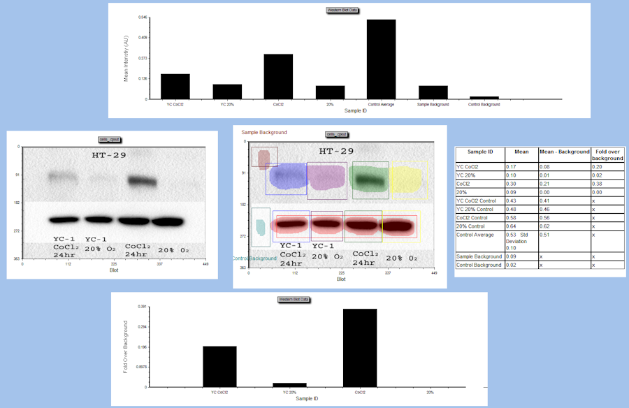

Figure 2: Overview of the CellProfiler pipeline using Galaxy tools.ĭetails: More details about the pipeline steps

Input and output file locations are set by Galaxy and can’t be set by the user.
#Cellprofiler intensity segmentation manual#
Parameters require manual input from the user whereas, in the stand-alone version, some modules can inherit parameter values from other modules.Parameters for some CellProfiler modules are limited/constrained compared to the stand-alone version, most notably:.Modules used by the pipeline aren’t available in Galaxy.The Galaxy tool currently uses CellProfiler 3.9. The pipeline was built with a different version of CellProfiler.Some pipelines created with stand-alone CellProfiler may not work with the Galaxy CellProfiler tool. The Galaxy CellProfiler Tool: toolshed.g2.bx.psu.edu/repos/bgruening/cp_cellprofiler/cp_cellprofiler/3.1.9+ galaxy0 tool takes two inputs: a CellProfiler pipeline and an image collection. Warning: Important information: CellProfiler in Galaxy Here we will link objects if they significantly overlap between the current and previous frames. Linking is done by matching objects and several criteria or matching rules are available. Tracking is done by first segmenting objects then linking objects between consecutive frames. To demonstrate how automatic tracking can be applied in such situations, this tutorial will track dividing nuclei in a short time-lapse recording of one mitosis of a syncytial blastoderm stage Drosophila embryo expressing a GFP-histone gene that labels chromatin. One of these challenges is the tracking of individual objects as it is often impossible to manually follow a large number of objects over many time points. However, automated time-lapse imaging can produce large amounts of data that can be challenging to process. Combining fluorescent markers with time-lapse imaging is a common approach to collect data on dynamic cellular processes such as cell division (e.g. Most biological processes are dynamic and observing them over time can provide valuable insights.


 0 kommentar(er)
